To develop effective treatments, we need to figure out the basics first.
Our research is crucial for uncovering this knowledge and ultimately finding a cure.
The second and third-place winners exemplify the diversity found in the competition.
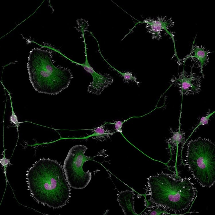
1st Place, Dr. Bruno Cisterna & Dr. Eric Vitriol, Medical College of Georgia at Augusta University, Department of Neuroscience & Regenerative Medicine. Differentiated mouse brain tumor cells (actin, microtubules, and nuclei), Super-Resolution, 40X (Objective Lens Magnification)
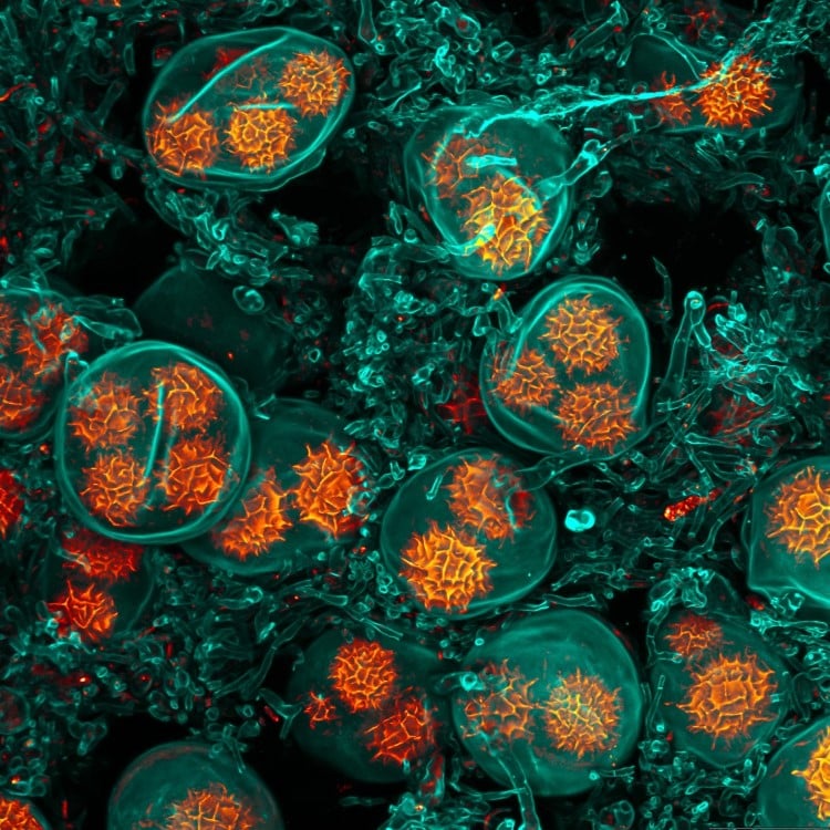
Third place was handed out to Chris Romaine for his fascinating look at a cannabis leaf with bulbous trichomes.
Sometimes, we overlook the tiny details of the world around us.
Here are the winners of the 2024 Nikon Small World Photomicrography Competition.

2nd Place, Dr. Marcel Clemens, Verona, Veneto, Italy. Electrical arc between a pin and a wireImage stacking for the pin and wire combined with long exposure for the electrical arcs, 10X (Objective Lens Magnification)
2nd Place, Dr. Marcel Clemens, Verona, Veneto, Italy.
Leaf of a cannabis plant.
The bulbous glands are trichomes.
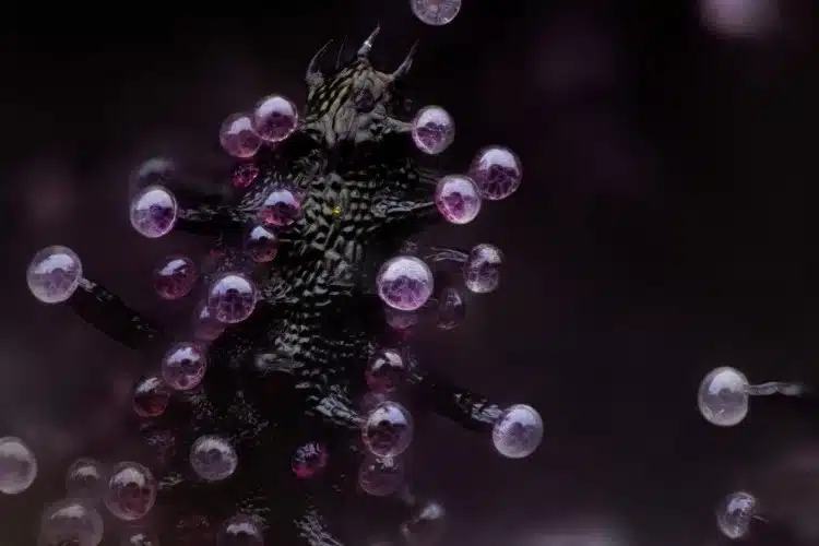
3rd Place, Chris Romaine, Kandid Kush, Port Townsend, Washington, USA. Leaf of a cannabis plant. The bulbous glands are trichomes. The bubbles inside are cannabinoid vesicles, Image Stacking, 20X (Objective Lens Magnification)
9th Place, John-Oliver Dum, Medienbunker Produktion, Bendorf, Rheinland Pfalz, Germany.
15th Place, Sebastien Malo, Saint Lys, Haute-Garonne, France.
Two water fleas (Daphnia sp.)
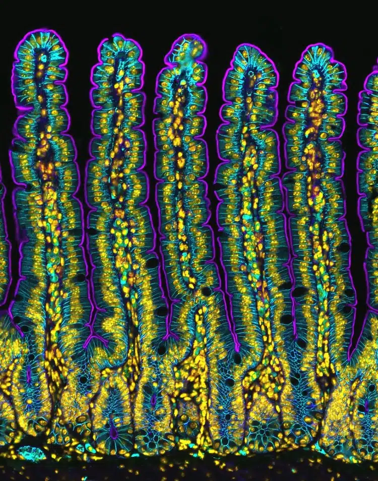
4th Place, Dr. Amy Engevik, Medical University of South Carolina Department of Regenerative Medicine & Cell Biology. Section of a small intestine of a mouse, Fluorescence10X (Objective Lens Magnification)
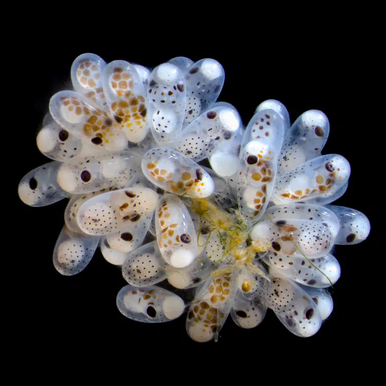
5th Place, Thomas Barlow & Connor Gibbons, Columbia University Department of Neurobiology and Behavior. Cluster of octopus (Octopus hummelincki) eggs, Darkfield, Stereomicroscopy, Focus Stacking, 3X (Objective Lens Magnification)
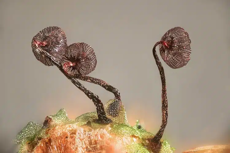
6th Place, Henri Koskinen, Helsinki University. Slime mold (Cribraria cancellata)Image Stacking, Polarized Light, Reflected Light, 10X (Objective Lens Magnification)
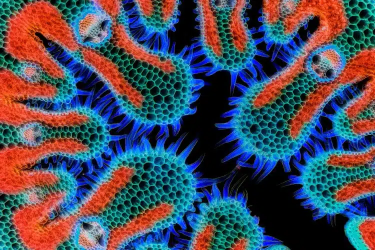
7th Place, Gerhard Vlcek, Maria Enzersdorf, Austria. Cross section of European beach grass (Ammophila arenaria) leaf, Brightfield, Image Stacking, 10X (Objective Lens Magnification)
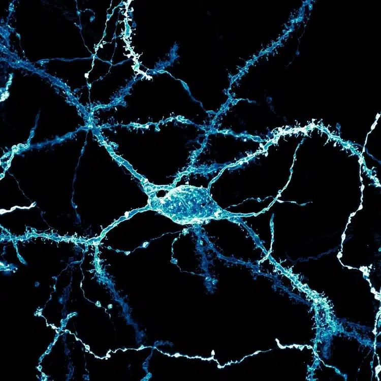
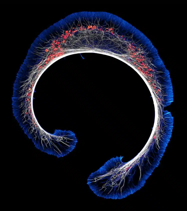
8th Place, Stephanie Huang, Victoria University of Wellington, School of Biological Sciences; School of PsychologyWellington, New Zealand. A neuron densely covered in dendritic spines from the striatum of an adult rat brain, Confocal, Deconvolution, Image Stacking, 60X (Objective Lens Magnification)
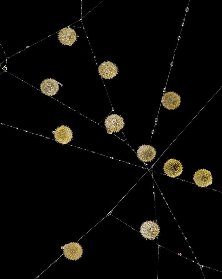
9th Place, John-Oliver Dum, Medienbunker Produktion, Bendorf, Rheinland Pfalz, Germany. Pollen in a garden spider (Araneus) web, Image Stacking, 20X (Objective Lens Magnification)
10th Place, Jan Martinek, Charles University Department of Experimental Plant Biology, Prague, Czech Republic. Spores of black truffle (Tuber melanosporum), Confocal, 63X (Objective Lens Magnification)
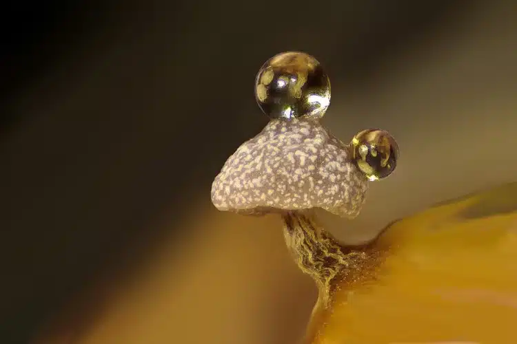
11th Place, Dr. Ferenc Halmos, Bánd, Veszprém, Hungary. Slime mold on a rotten twig with water droplets, Image Stacking, 0.7X – 4.5X (Objective Lens Magnification)
12th Place, Daniel Knop, Oberzent-Airlenbach, Hessen, Germany. Wing scales of a butterfly (Papilio ulysses) on a medical syringe needle, Image Stacking, 20X (Objective Lens Magnification)
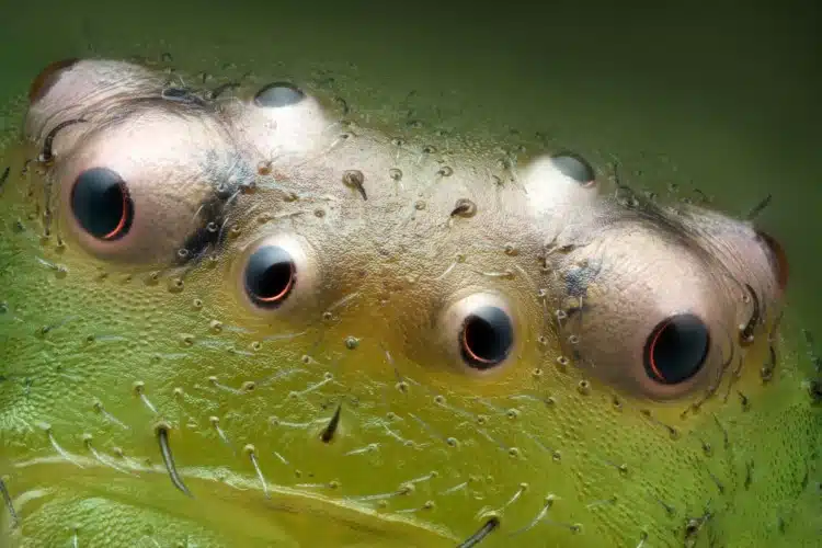
13th Place, Paweł Błachowicz, Bedlno, Świętokrzyskie, Poland. Eyes of green crab spider (Diaea dorsata), Image Stacking, Reflected Light, 20X (Objective Lens Magnification)
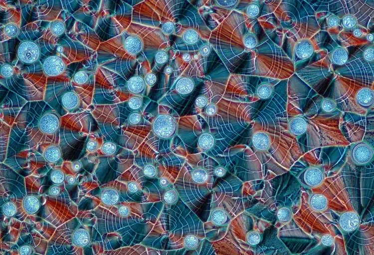
14th Place, Marek Miś, Marek Miś Photography, Suwalki, Podlaskie, Poland. Recrystallized mixture of hydroquinone and myoinositol, Polarized Light, 10X (Objective Lens Magnification)
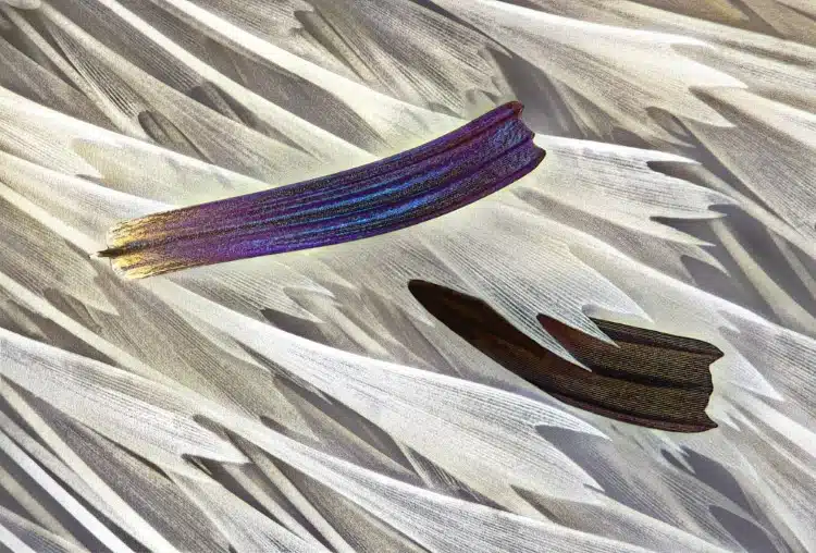
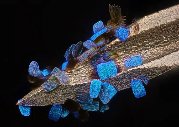
15th Place, Sébastien Malo, Saint Lys, Haute-Garonne, France. Isolated scales on Madagascan sunset moth wing (Chrysiridia ripheus), Darkfield, Image Stacking, Reflected Light, 40X (Objective Lens Magnification)
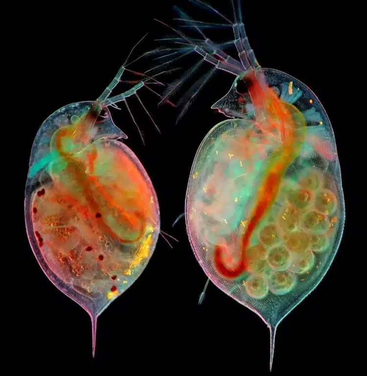
16th Place, Marek Miś, Marek Miś Photography, Suwalki, Podlaskie, Poland. Two water fleas (Daphnia sp.) with embryos (left) and eggs (right), Darkfield, Polarized Light, 10X (Objective Lens Magnification)
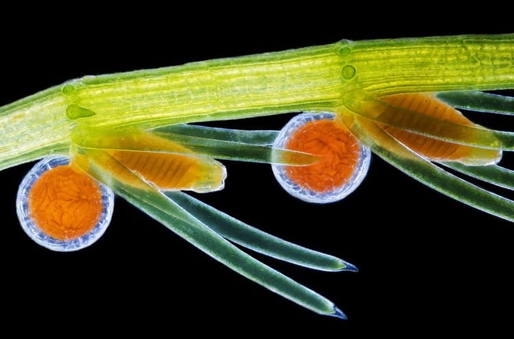
17th Place, Dr. Frantisek Bednar, Svosov, Zilinsky, Slovak Republic. Stonewort algae (Chara virgata) reproductive organs – oogonia (female organs) and antheridia (male organs), Darkfield, 4X (Objective Lens Magnification)
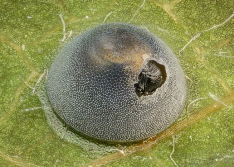
18th Place, Alison Pollack, San Anselmo, California, USA. An insect egg parasitized by a wasp, Image Stacking, Reflected Light, 10X (Objective Lens Magnification)
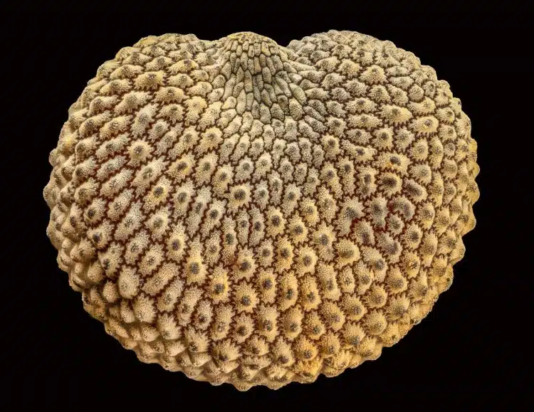
19th Place, Alison Pollack, San Anselmo, California, USA. Seed of a Silene plant, Image Stacking, Reflected Light, 10X (Objective Lens Magnification)
20th Place, Dr. Bruno Cisterna & Dr. Eric Vitriol, Medical College of Georgia at Augusta UniversityDepartment of Neuroscience & Regenerative Medicine. Early stage of mouse glioblastoma cell differentiation (actin, microtubules, and mitochondria), Super-Resolution, 100X (Objective Lens Magnification)
