I entered the competition as a way to showcase the complexity of retinal microcirculation.
As usual, all of the top 20 images highlight the artistry of science.
With a wide variety of subjects and photomicrography techniques, the winning scientists and researchers are certainly inspirational.
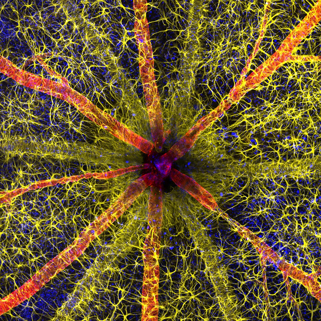
1st Place, Hassanain Qambari & Jayden Dickson., The Lions Eye Institute Department of Physiology & Pharmacology, Perth, Western Australia, Australia. Rodent optic nerve head showing astrocytes (yellow), contractile proteins (red) and retinal vasculature (green). Confocal, Fluorescence, Image Stacking, 20X (Objective Lens Magnification)
These are the winners of the 2023 Nikon Small World Photomicrography Competition.
2nd Place, Ole Bielfeldt, Macrofying, Cologne, North Rhine-Westphalia, Germany.
Matchstick igniting by the friction surface of the box.
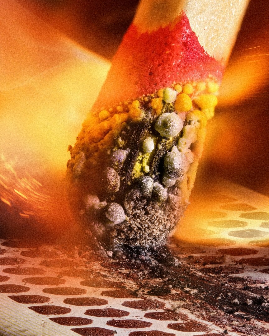
2nd Place, Ole Bielfeldt, Macrofying, Cologne, North Rhine-Westphalia, Germany. Matchstick igniting by the friction surface of the box. Brightfield, Image Stacking, 2.5X (Objective Lens Magnification)
Venomous fangs of a small tarantula.
Slime mold (Comatricha nigra) showing capillitial fibers through its translucent peridium.
Blood and lymphatic vasculatures in the ear skin of an adult mouse.
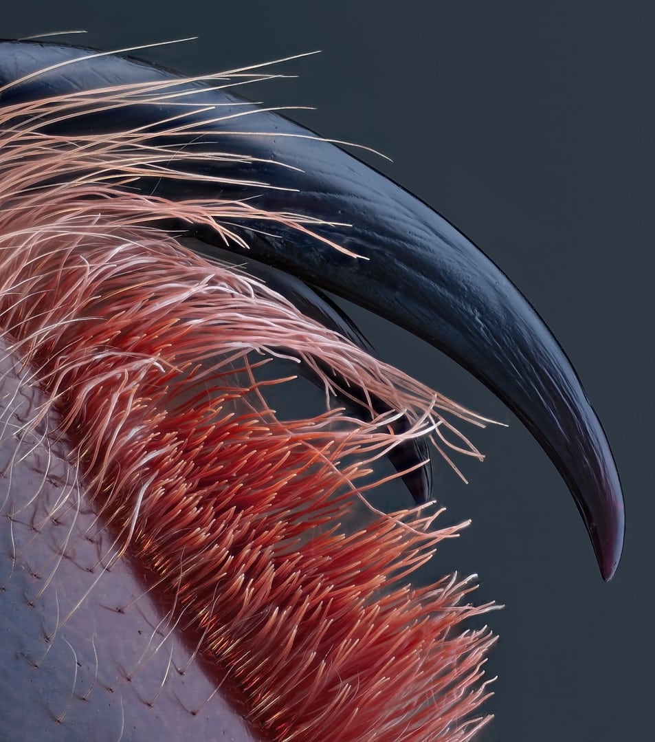
4th Place, John-Oliver Dum, Medienbunker Produktion, Bendorf, Rheinland-Pfalz, Germany. Venomous fangs of a small tarantula. Image Stacking, 10X (Objective Lens Magnification)
Confocal 10X, (Objective Lens Magnification)
12th Place, Sherif Abdallah Ahmed.
Faculty of Science, Tanta University, Department of Zoology, Tanta, Egypt, Arab Republic.
Cuckoo wasp standing on a flower.
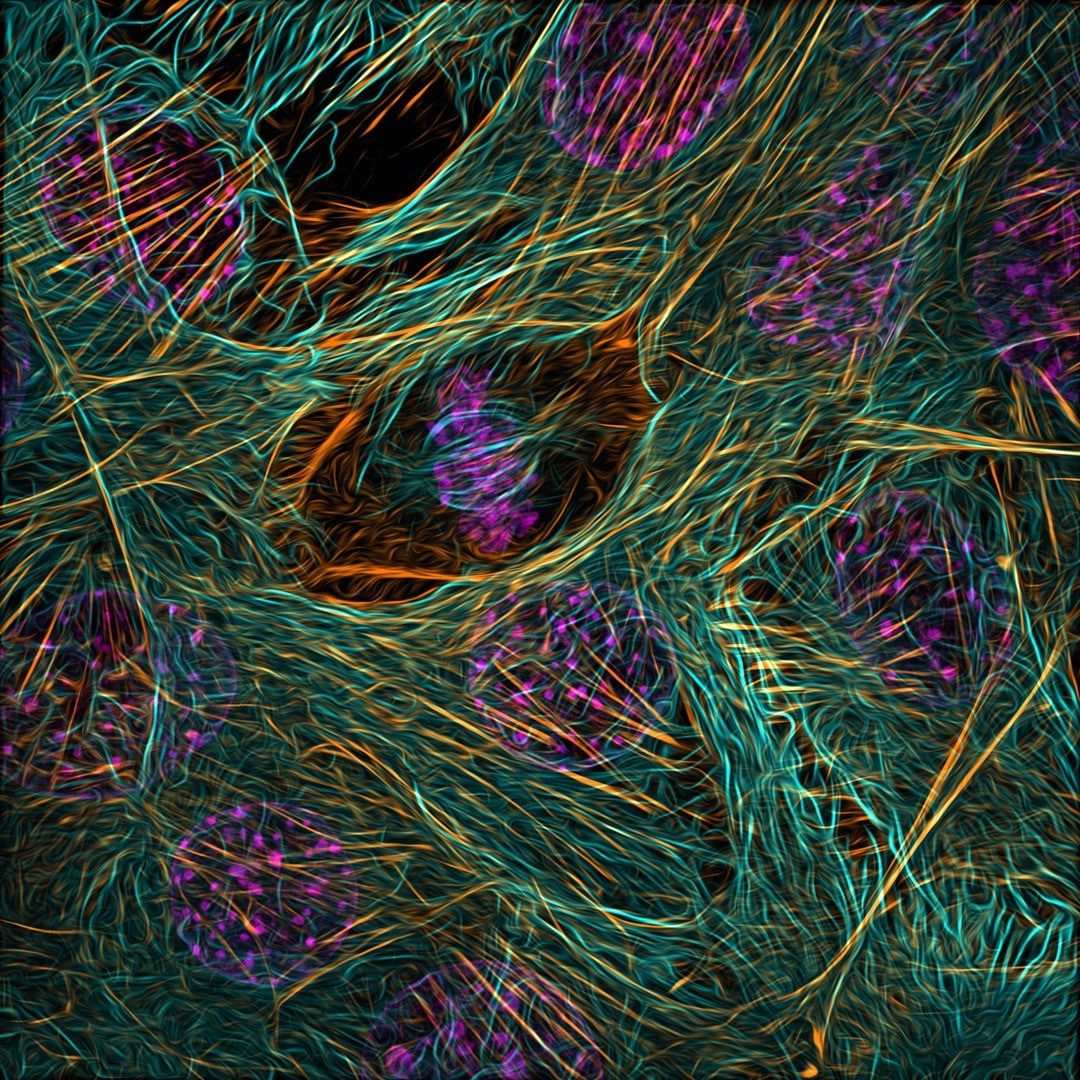
9th Place, Vaibhav Deshmukh, Baylor College of Medicine, Department of Molecular Physiology and Biophysics, Houston, Texas, USA. Cytoskeleton of a dividing myoblast; tubulin (cyan), F-actin (orange) and nucleus (magenta). Fluorescence, Structured Illumination Microscopy (SIM), 63X (Objective Lens Magnification)
Sunflower pollen on an acupuncture needle.
3rd Place, Malgorzata Lisowska, Independent, Value Based Healthcare Consultant, Warsaw, Mazowieckie, Poland.
Motor neurons grown in microfluidic gear for separation of cell bodies (top) and axons (bottom).
11th Place, Dr. Diego García, Universidad Complutense de Madrid, Real Sociedad Española de Física, Madrid, Spain. Crystallized sugar syrup. Polarized Light, 25X (Objective Lens Magnification)
Green microtubules; Red growth cones (actin).
Auto-fluorescing defensive hairs covering the leaf surface of Eleagnus angustifolia exposed to UV light.
Fluorescence, Image Stacking, 10X (Objective Lens Magnification)
15th Place, Dr. Pichaya Lertvilai.
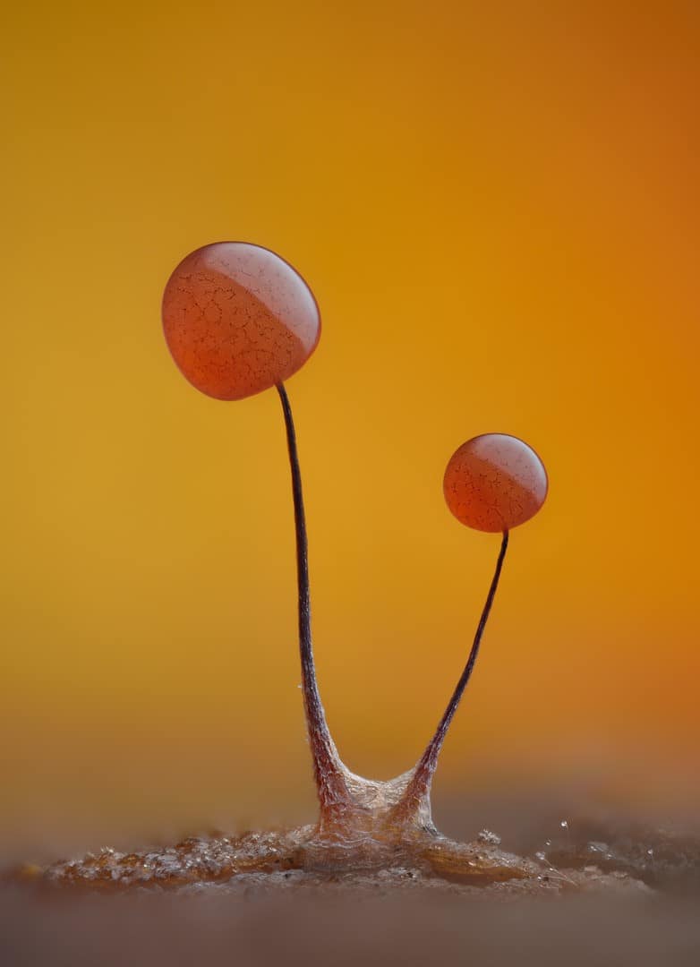
6th Place, Timothy Boomer, WildMacro.com, Vacaville, California, USA. Slime mold (Comatricha nigra) showing capillitial fibers through its translucent peridium. Image Stacking, 10X (Objective Lens Magnification)
UC San Diego, Scripps Institution of Oceanography, La Jolla, California, USA.
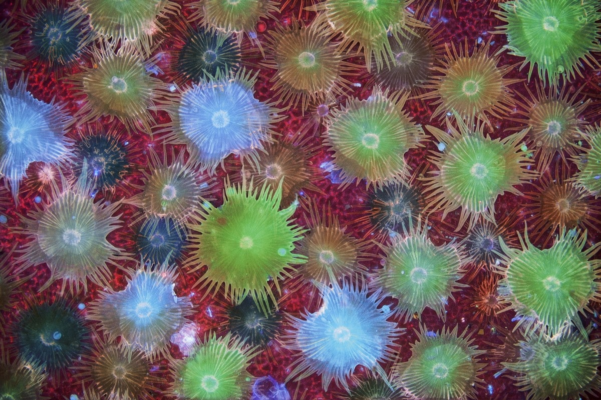
Fluorescent image of an Acropora sp.
showing individual polyps with symbiotic zooxanthellae.
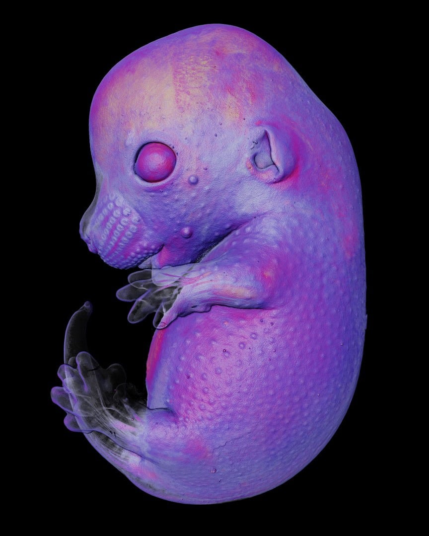
7th Place, Dr. Grigorii Timin & Dr. Michel Milinkovitch, University of Geneva, Department of Genetics and Evolution, Geneva, Switzerland. Mouse embryo. Light Sheet, 4X (Objective Lens Magnification)
Darkfield, Fluorescence, Image Stacking, 5X (Objective Lens Magnification)
Related Articles:
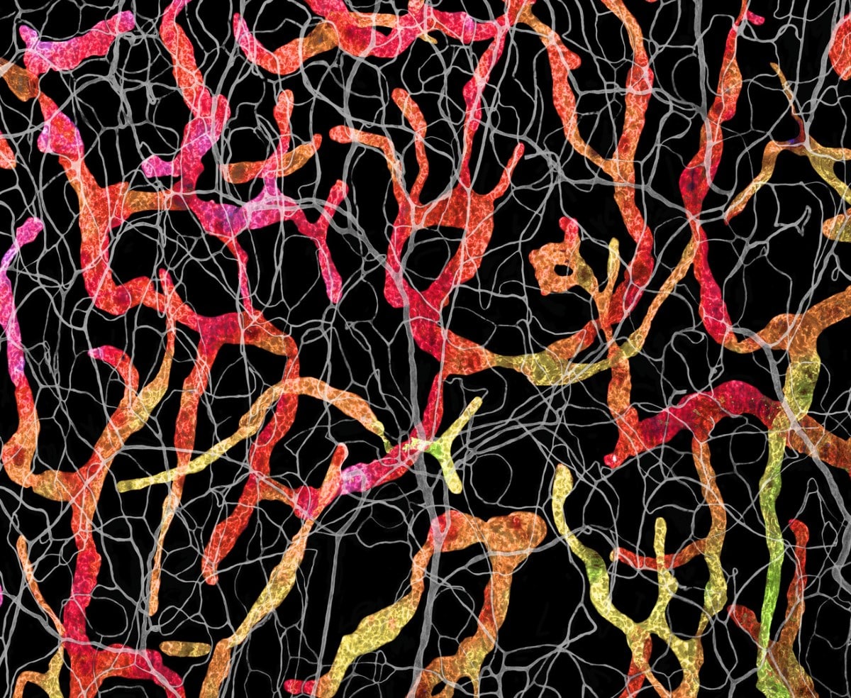
13th Place, Satu Paavonsalo & Dr. Sinem Karaman, University of Helsinki, Individualized Drug Therapy Research Program, Faculty of Medicine, Helsinki, Finland. Blood and lymphatic vasculatures in the ear skin of an adult mouse. Confocal 10X, (Objective Lens Magnification)
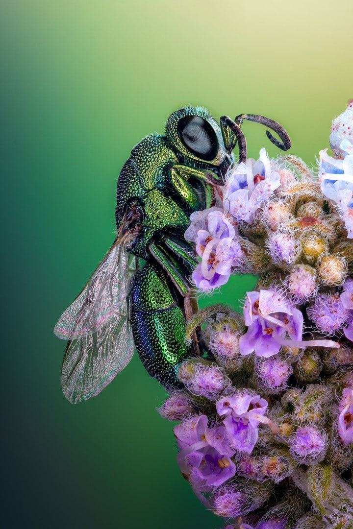
12th Place, Sherif Abdallah Ahmed. Faculty of Science, Tanta University, Department of Zoology, Tanta, Egypt, Arab Republic. “Cuckoo wasp” standing on a flower. Image Stacking, 4X (Objective Lens Magnification)
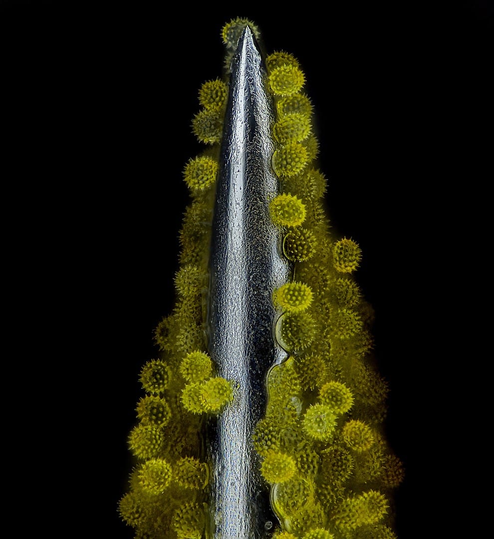
14th Place, John-Oliver Dum, Medienbunker Produktion, Bendorf, Rheinland-Pfalz, Germany. Sunflower pollen on an acupuncture needle. Image Stacking, 40X (Objective Lens Magnification)

3rd Place, Malgorzata Lisowska, Independent, Value Based Healthcare Consultant, Warsaw, Mazowieckie, Poland. Breast cancer cells. Brightfield, Image Stacking, 40X (Objective Lens Magnification)
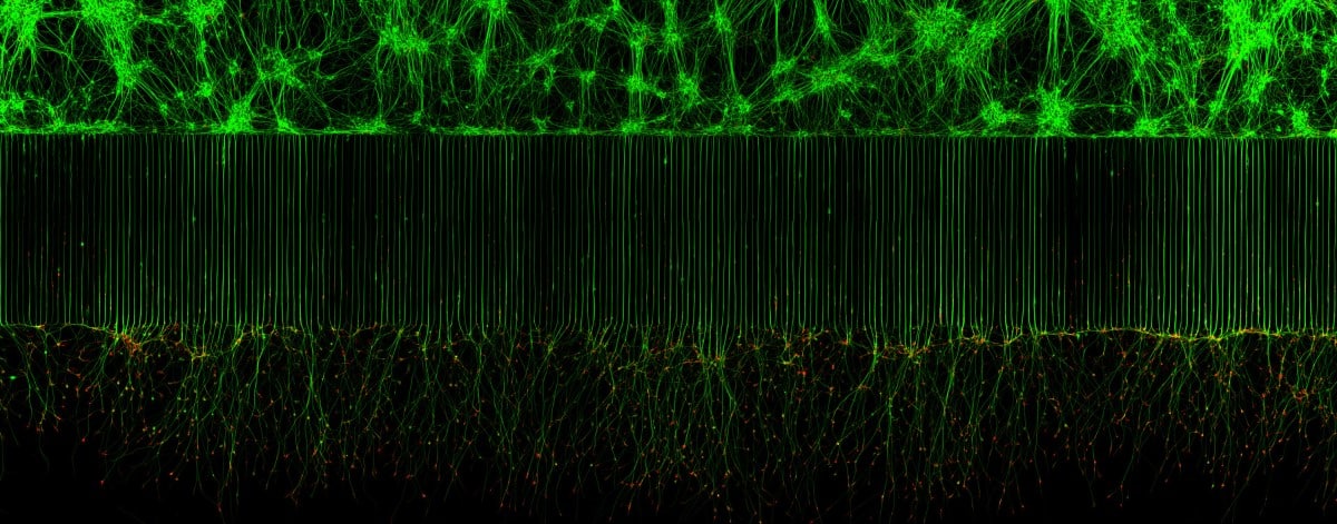
10th Place, Melinda Beccari & Dr. Don W. Cleveland, UC San Diego, Department of Cellular and Molecular Medicine, La Jolla, California, USA. Motor neurons grown in microfluidic device for separation of cell bodies (top) and axons (bottom). Green – microtubules; Red – growth cones (actin). Confocal, Fluorescence, 20X (Objective Lens Magnification)
8th Place, Stefan Eberhard, University of Georgia, Athens, Georgia, USA. Caffeine crystals. Polarized Light, 25X (Objective Lens Magnification)
5th Place, Dr. David Maitland, Feltwell, Norfolk, United Kingdom. Auto-fluorescing defensive hairs covering the leaf surface of Eleagnus angustifolia exposed to UV light. Fluorescence, Image Stacking, 10X (Objective Lens Magnification)

15th Place, Dr. Pichaya Lertvilai. UC San Diego, Scripps Institution of Oceanography, La Jolla, California, USA. Fluorescent image of an Acropora sp. showing individual polyps with symbiotic zooxanthellae. Darkfield, Fluorescence, Image Stacking, 5X (Objective Lens Magnification)
