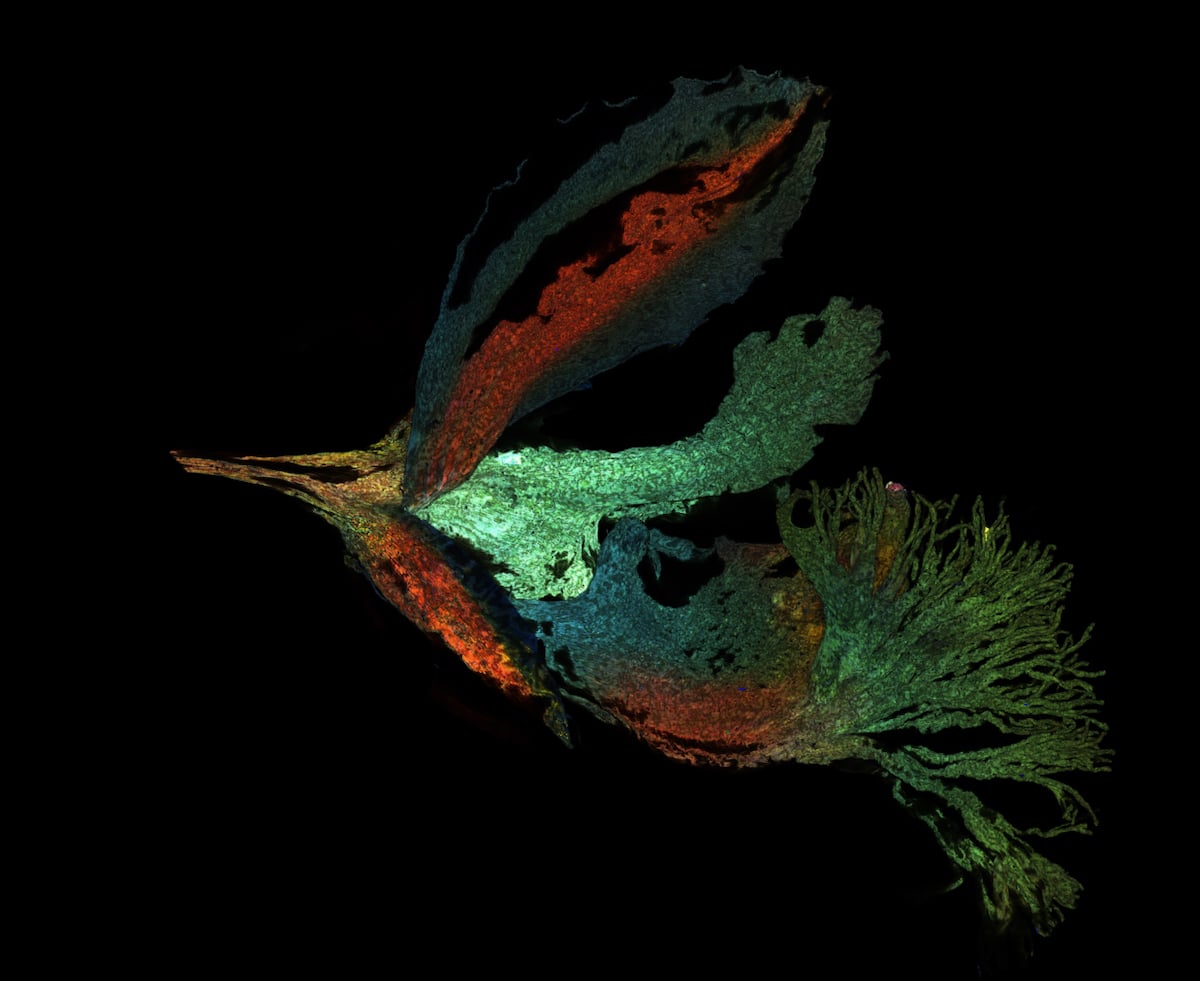
Global winner Jan Martinek (Czech Republic).Arabidopsis thaliana flower with pollen tubes growing through the pistil.
Nearly 800 images were submitted to theOlympus Image of the Year Award 2021.
The winning images create a beautiful image gallery showcasing an incredible level of talent and technique at the microscope.
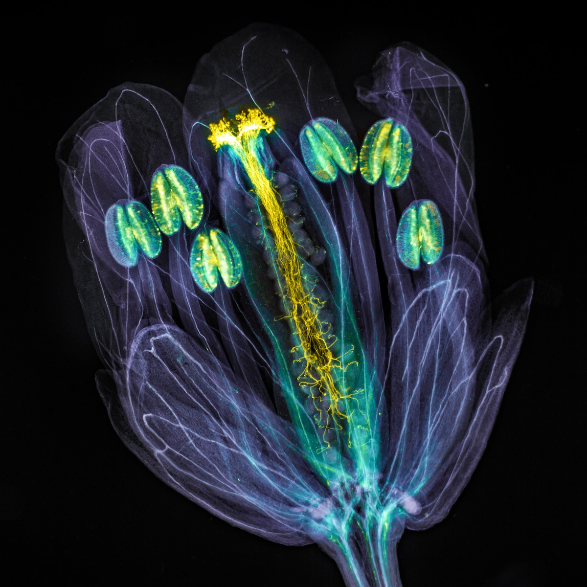
Global winner – Jan Martinek (Czech Republic).“Arabidopsis thaliana flower with pollen tubes growing through the pistil. The flower tissues were chemically cleared to become transparent, while the pollen tubes were stained with aniline blue (yellow fluorescence) in order to be seen.”
All of the winners received Olympus microscopes to further their scientific and artistic pursuits.
Olympus Image of the Year Award 2021 celebrates the best microscopy across the globe.
Cell walls (in cyan) were stained live with calcofluor white.
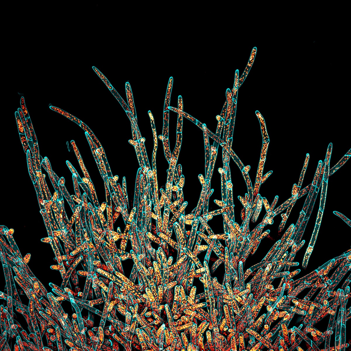
Regional Winner (Americas) – Ivan Radin (USA).“A maximum projection of the deconvolved Z-stack of moss Physcomitrium patens protonemal cells. Cell walls (in cyan) were stained live with calcofluor white. Chloroplasts autofluorescence is in Fall LUT.”
Chloroplasts autofluorescence is in Fall LUT.
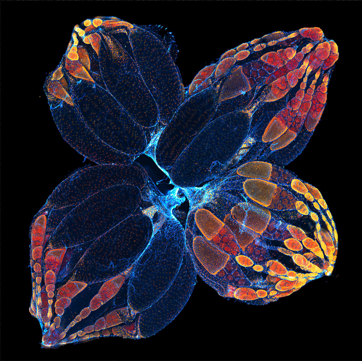
Regional winner (Asia-Pacific) Daniel Han (Australia).Fern sori capsules with spores bursting out.
Honorable mention Yayun Wang (China).Mouse brain GABA neurons.
Regional winner (Asia-Pacific) – Daniel Han (Australia).“Fern sori capsules with spores bursting out. Captured using Z-stacking.”
Over 800 images taken with microscopes from 49 countries were submitted to the contest.
Honorable mention Mingyue Jia (China).Autofluorescence image of Siberian polygala.
Captured using confocal microscopy.
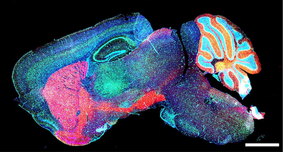
Honorable mention – Yayun Wang (China).“Mouse brain GABA neurons.”
Rendered using maximum projection.
Honorable mention Yujun Chen (USA).Ovaries of the fruit fly.
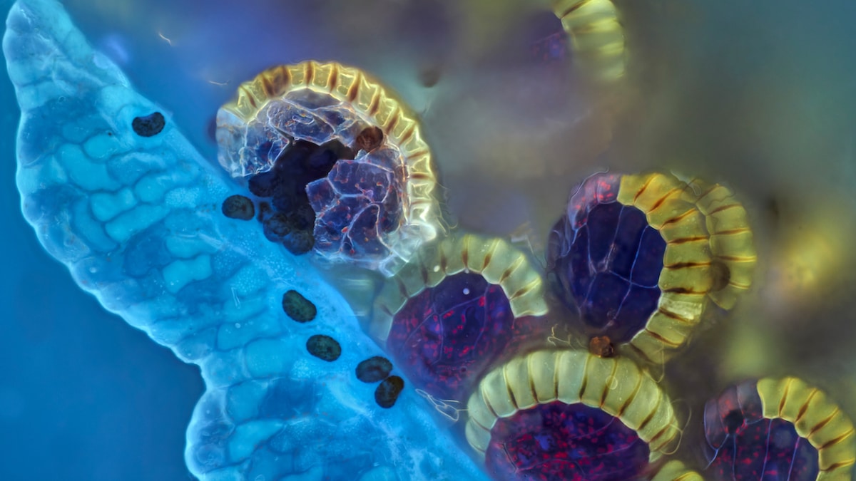
One cell typically carries one nucleus.
Honorable mention – Mingyue Jia (China).“Autofluorescence image of Siberian polygala. Captured using confocal microscopy. Rendered using maximum projection.”
In contrast, as seen here, an arbuscular mycorrhizal (AM) fungal cell carries hundreds of nuclei.
Stained with Congo red.
Imaged using a 10X (0.45 NA) objective.
Honorable mention – Yujun Chen (USA).“Ovaries of the fruit fly.”
Overall and regional winners were awarded and several images were highlighted as honorable mentions.
The hairs are silhouetted against the leafs red-fluorescent, chlorophyll-packed cells.
Honorable mention LayraCintron-Rivera (USA).The developing nervous system of an embryonic zebrafish.
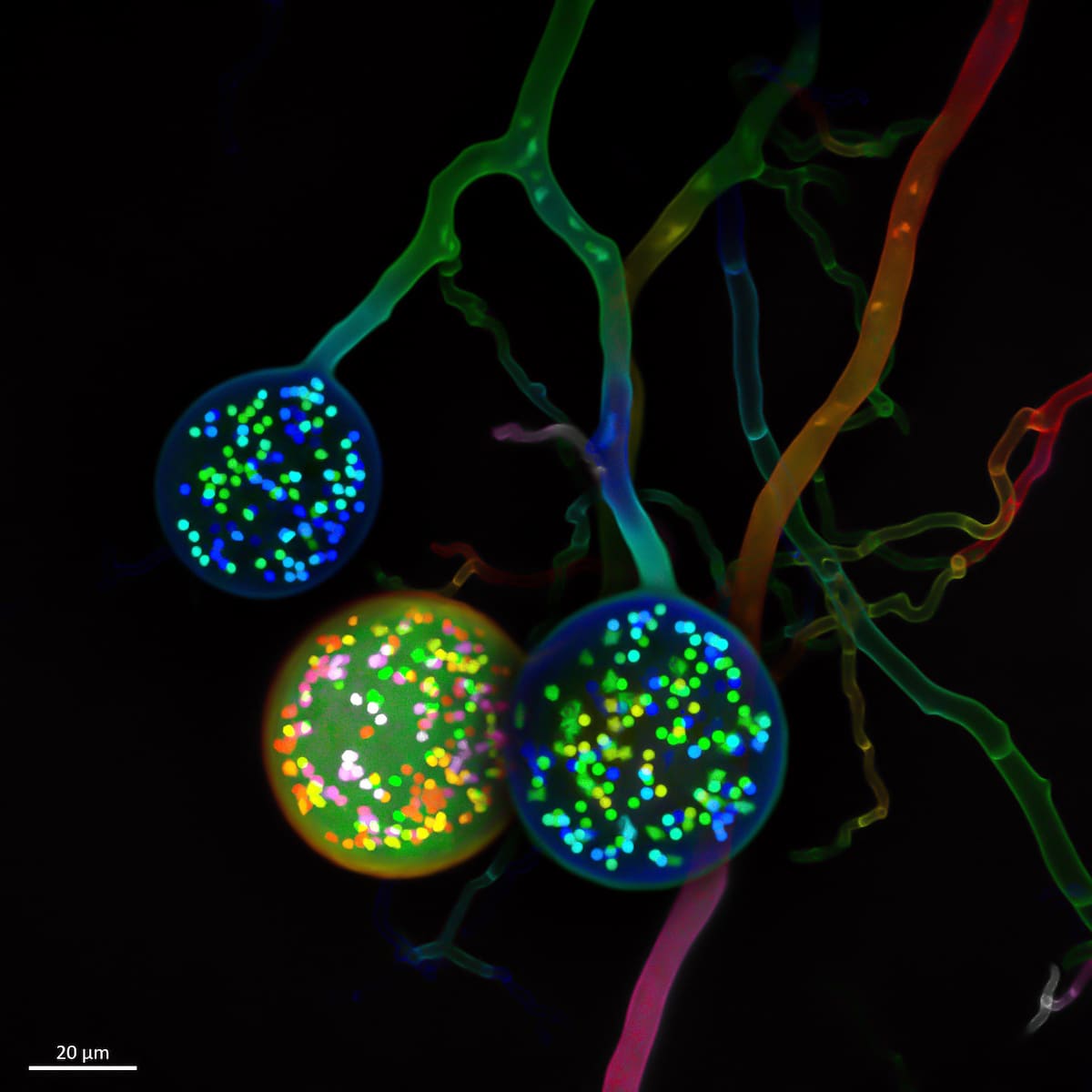
Regional winner (Europe, the Middle East, and Africa) – Vasilis Kokkoris (Netherlands).“Multinucleate spores of a soil fungus. One cell typically carries one nucleus. In contrast, as seen here, an arbuscular mycorrhizal (AM) fungal cell carries hundreds of nuclei.”
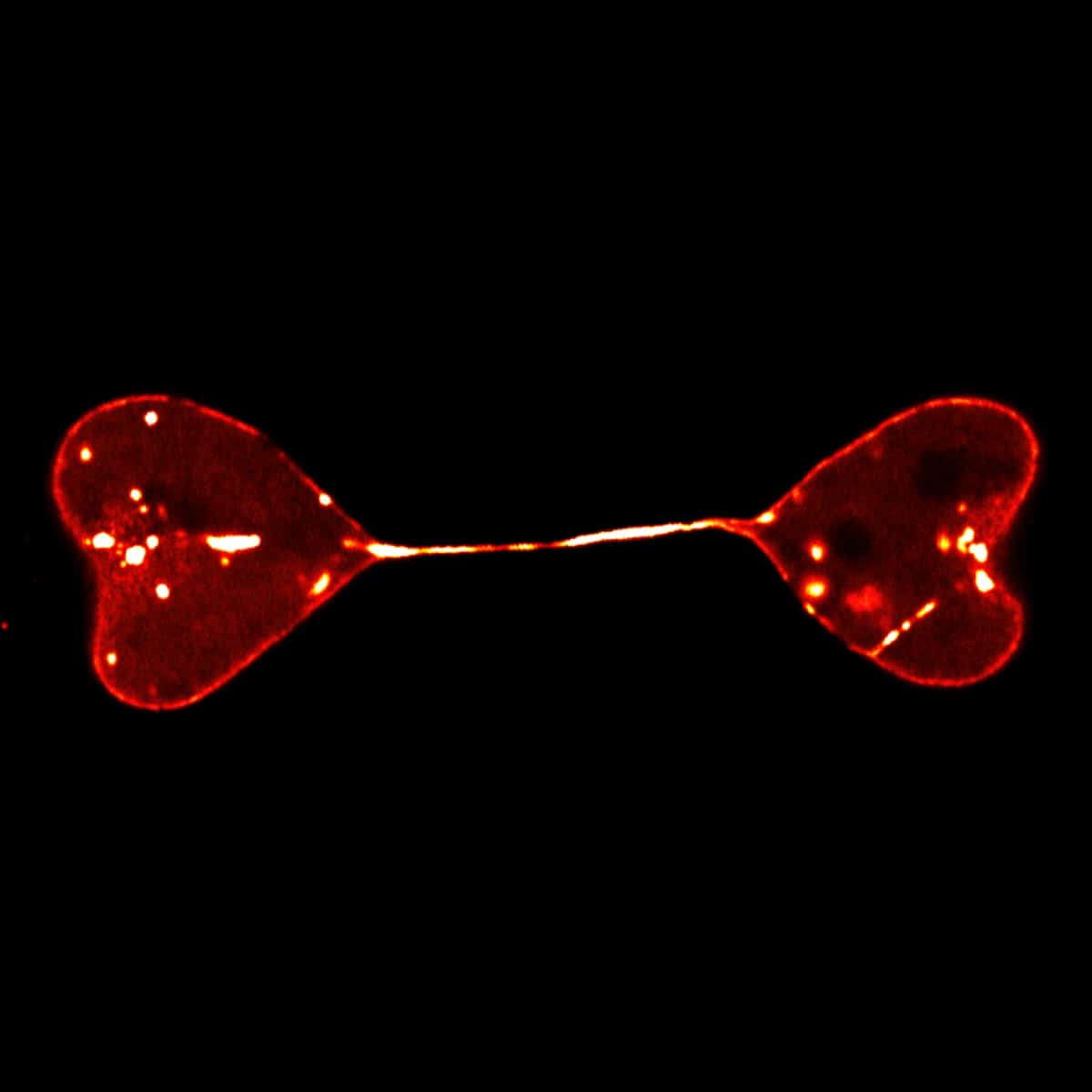
Honorable mention Di Lu (China).Semi-separated nuclei of two cells form a heart-to-heart shape.
The nuclei were labeled by lamin.
Related Articles:
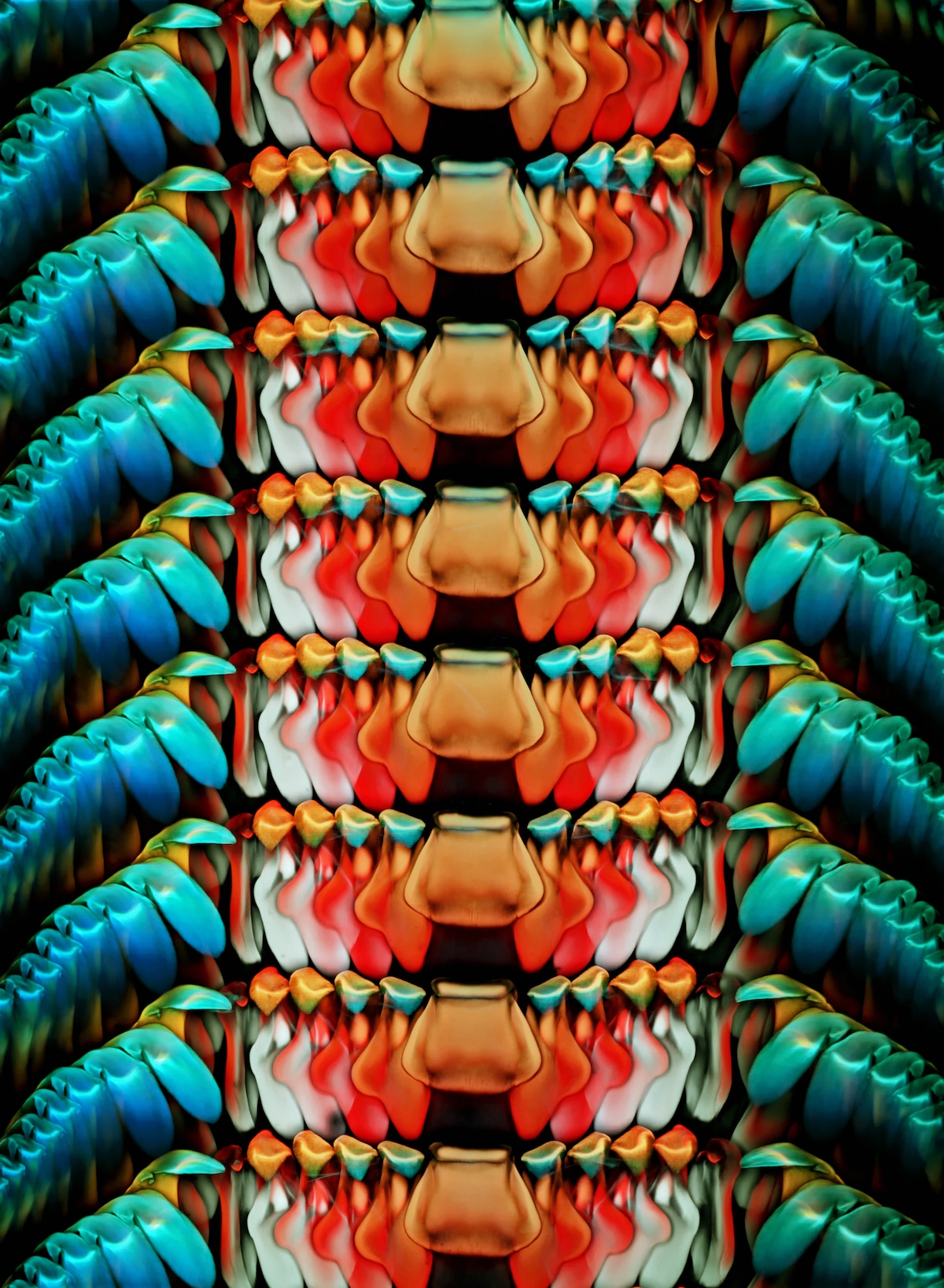
Honorable mention – Igor Siwanowiczfrom (USA).“Rasping tongue, or radula, of an Astraea conehead snail, Astraea tecta. Stained with Congo red. Imaged using a 10X (0.45 NA) objective. Depth color-coded projection.”
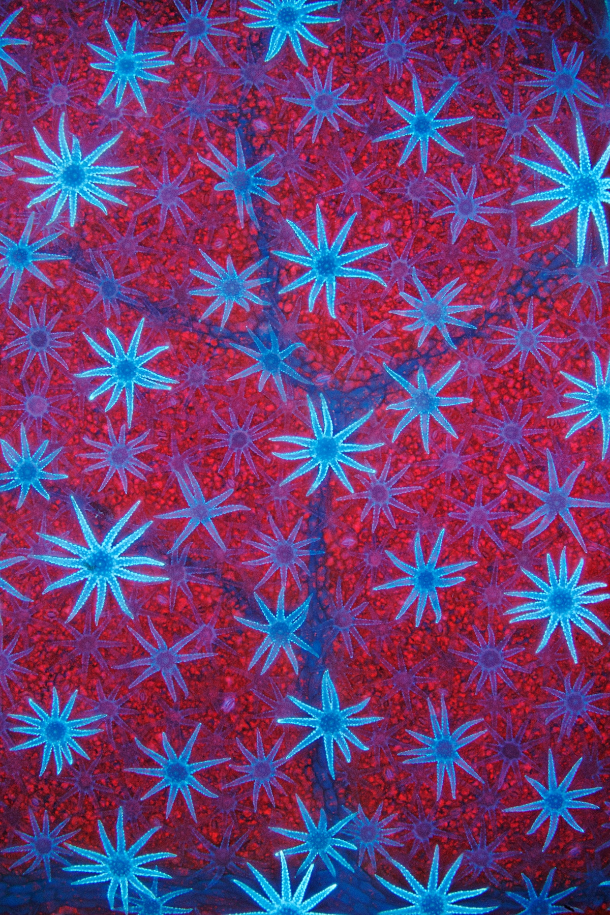
Honorable mention – David Maitland (UK).“Blue autofluorescent, star-shaped defensive hairs cover the surface of a Deutzia leaf. The hairs are silhouetted against the leaf’s red-fluorescent, chlorophyll-packed cells.”
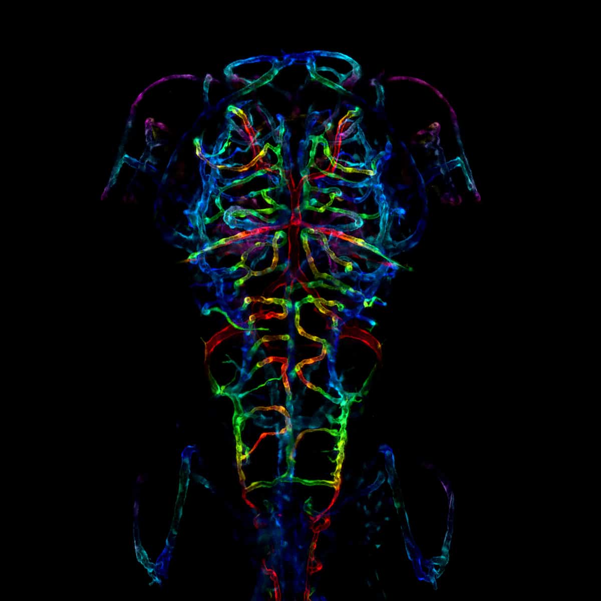
Honorable mention – LayraCintron-Rivera (USA).“The developing nervous system of an embryonic zebrafish. Specifically, the image is a color-coded projection of the axonal projections of a zebrafish fixed six days after fertilization.”
Honorable mention – Di Lu (China).“Semi-separated nuclei of two cells form a heart-to-heart shape. The nuclei were labeled by lamin.”
